What is a bacteriophage?
Bacterio refers to “bacteria” and phage to “eat”, thus bacteriophages are viruses that grow on bacteria by attaching to the bacterial cell wall to penetrate and inject their genetic material, which degrades the bacteria’s genetic material. The viral DNA/RNA replicates within the bacteria, until the bacteria wall lyses and more phages are released.
Phages were discovered to be antimicrobial agents for treating bacterial infections in the late 1800’s and early 1900’s. Antimicrobial resistance presents one of the greatest challenges of modern medicine and the specificity of phages to disease-causing bacterial strains make them particularly attractive alternatives to traditional antibiotics.

Left image: Bacteriophage structure (a) and binding to bacteria. Right image: Nobrega, F.L., Vlot, M., de Jonge, P.A. et al. Targeting mechanisms of tailed bacteriophages. Nat Rev Microbiol 16, 760–773 (2018). AMI IMAGES/SCIENCE SOURCE
Bacterial plaquing
For clinical analysis of bacteriophage treatment, a simple and reproducible assay is required to quantify the levels of an engineered bacteriophage in a sample. This is achieved by screening for bacteriophage activity using a bacterial plaquing assay. A plaque is a clear area on an opaque field of target bacteria growing on an agar plate. The clear area indicates the bacteriophage-mediated antibacterial activity. These plaques can be enumerated and correlate with the number of bacteriophages in the sample, permitting easy quantification.
Synexa R&D proof-of-concept
Synexa’s R&D scientists used a specific E.coli targeting bacteriophage and commercial E.coli culture to demonstrate the utility of the bacterial plaquing assay. The assay procedure in brief involved mixing the E.coli culture and titrated bacteriophage solution prior to pouring into an agar plate. After incubation of 12-18 hours at 37°C, the plaques could be counted on each plate, corresponding to a specific bacteriophage dilution.
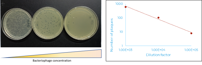
Left image: Plaque number was directly proportional to the dilution factor. Right image: the graph shows as bacteriophage concentration in the sample was decreased, plaque number decreased.
The results from our proof-of-concept work demonstrated the linearity in the relationship between plaque number and the concentration of bacteriophage in the sample.
For those currently investigating a bacteriophage therapy, this assay design can be easily modified to fit the target bacteria of interest and validated for the quantitative assessment of bacteriophage titres in a biological sample.
If you have any questions or would like to learn more about our bacteriophage-related assays, please contact us here