Tissue Imaging & Cell Solutions
Advanced multi-dimensional imaging capabilities tailored for drug development
Precise Target
Insights
Synexa’s specialised tissue and cell imaging services allow for the interrogation of multiple targets in situ. Leveraging advanced technologies, we provide comprehensive and precise insights into spatial relationships in cells and tissues, helping to achieve:
- More accurate target identification
- Selection of optimal drug candidates
- Development of robust biomarkers
Understanding Spatial Context
Spatial context is essential to understanding how cells organise and interact across the tissue landscape to drive disease progression and respond to therapy. New potential drugs may exhibit high efficacy in vitro or in animal models but fail in clinical trials due to the inability to accurately predict their behaviour within the complex spatial organisation of human tissues. The lack of spatial information often leads to a poor understanding of the drug’s mechanism of action, potential off-target effects and its interaction with different cell types within a tissue.
Our services empower researchers to gain a deeper understanding of tissue microenvironments and cellular behaviours, allowing for the precise understanding of therapeutic drug monitoring and efficacy.
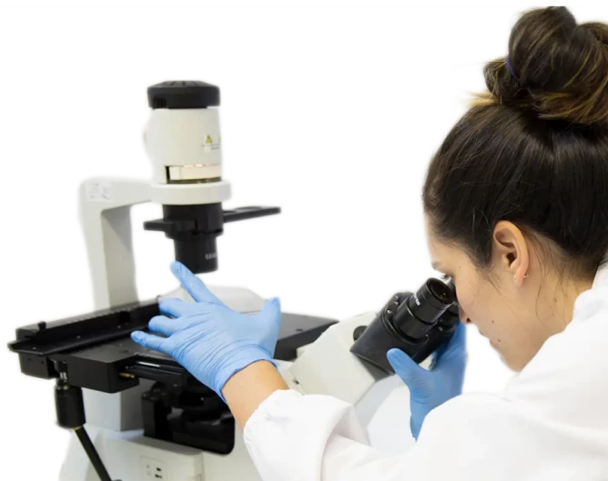
Tissue Imaging Services
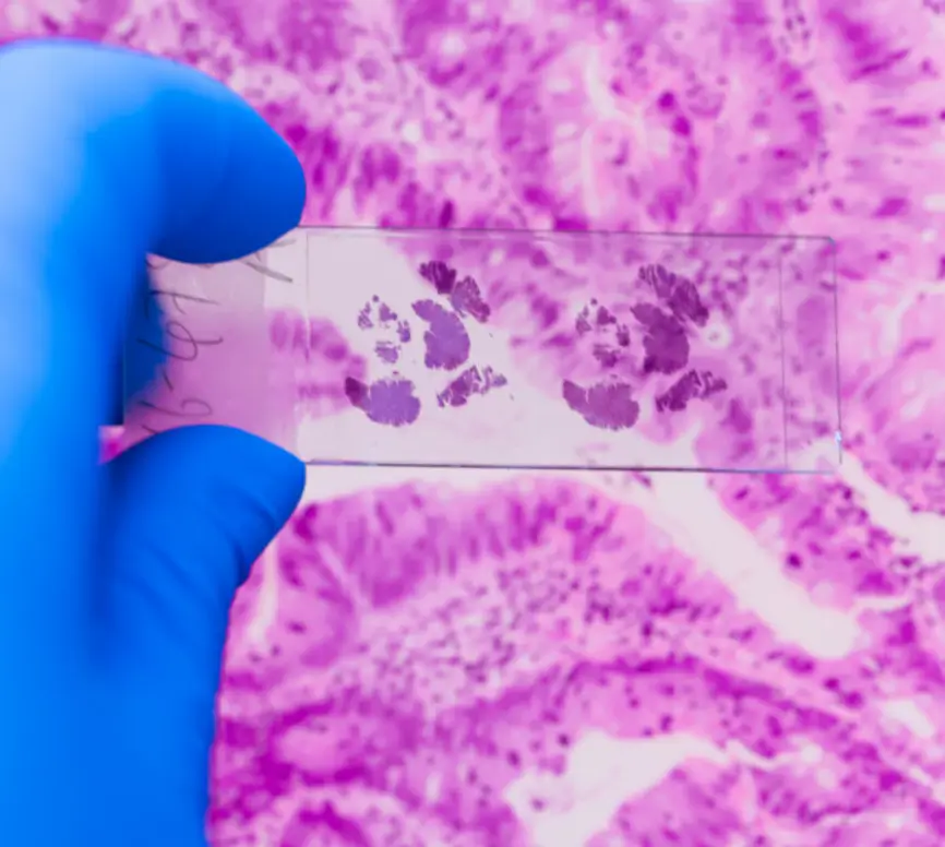
Whole Tissue Analysis
Spatial biology analysis provides a nuanced understanding of cellular dynamics and responses within a spatial context. We employ a synergistic approach that integrates traditional hematoxylin and eosin staining techniques with advanced and customisable multi-plex (4 and 8 plex) immunofluorescence panels (mIF). This allows for the simultaneous detection of multiple biomarkers within a single tissue section.
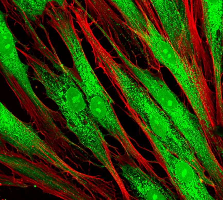
Super Resolution Microscopy (SRM)
Super Resolution Imaging enables the analysis of single molecules and events within cells, allowing for the observation of fluorescence images of subcellular organelles beyond the diffraction limit.
- Observe organelle interactions in living cells, find possible targets for treating diseases and observe the influence of drugs on the target
- Unveil critical insights into cellular processes and mechanisms such as intracellular cargo/drug trafficking and delivery, drug induced mitochondrial and microtubule dysfunction and DNA damage in response to drug therapy
- Identify specific biological processes and mechanisms for preclinical applications
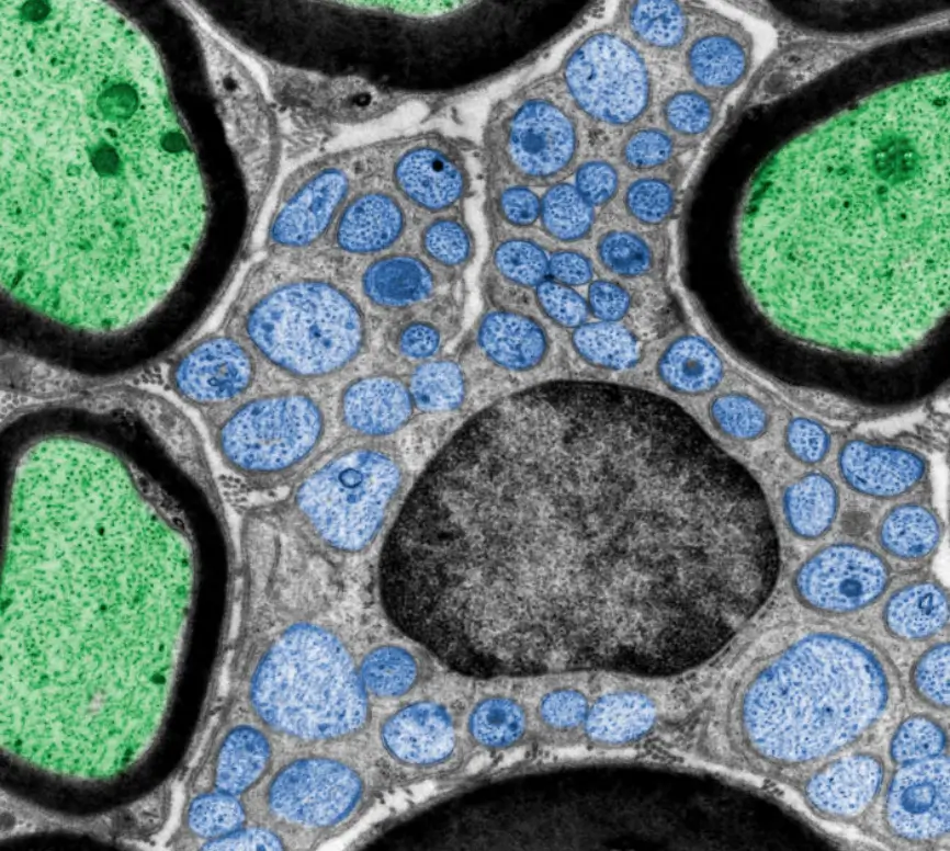
Correlative Light and Electron Microscopy (CLEM)
While fluorescence imaging allows identification of specific entities based on the labelling with fluorophores, the unlabeled constituents of the samples remain invisible. Correlative Light and Electron Microscopy (CLEM) enables ultrastructural analysis by combining the specificity of fluorescent labelling and the sub-nanometer resolution of electron microscopy.
- CLEM provides different views of the same sample, facilitating analysis of rare structures or cells and revealing ultrastructural context
- Determine whether a particular membrane-associated protein lies inside or outside a cell or organelle; select specific fluorescently stained cells for detailed structural interrogation; and probe the molecular stoichiometry and heterogeneity of subcellular features
- CLEM has largely been used in the field of neuroscience allowing insight into neuronal and synaptic functions of the brain and nervous system
Robust and Proven Platforms
Our technology platforms are robust, reliable and reputable in the clinical field and provide comprehensive and detailed data readouts for your application.
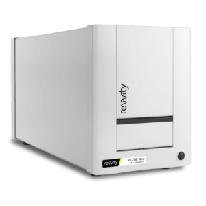
VICTOR® Nivo™
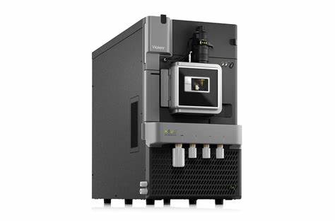
Waters T-QS LC-MS/MS

Improving the quality of human health
Contact us
Discuss biomarker and bioanalytical solutions to accelerate your therapeutic pipeline